
Chronic Venous Insufficiency: Evaluation with Doppler Ultrasound
- Dr. Segnini

- Jan 9
- 2 min read

A Dynamic Problem
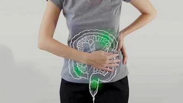
Chronic venous insufficiency (CVI) is a dysfunction of the superficial and/or deep venous system of the lower extremities, where the valves become incompetent, preventing the effective return of blood to the heart. This causes reflux, venous hypertension, and its clinical manifestations: varicose veins, edema, skin changes (ochre dermatitis, lipodermatosclerosis ), and, in advanced stages, venous ulcers. Doppler ultrasound is the gold standard for its diagnosis and treatment planning.
Doppler Ultrasound ( Venous Duplex ):
Unlike the study of thrombosis, here the focus is on function and hemodynamics .
Exact Anatomical Map: Identifies all saphenous veins (great and small), their collaterals and perforators, and the deep venous system.
Quantifies Reflux: Measures the time and direction of blood flow after a stimulus, determining valvular incompetence.
Locate the Leakage Point: Accurately identify the points where reflux passes from the deep system to the superficial system (perforating vein insufficiency) or where reflux begins in the saphenous system .
Therapeutic Guide: Essential for planning procedures such as endovenous laser or radiofrequency ablation, ultrasound-guided sclerotherapy, or surgery.
Protocol and Key Ultrasound Findings:
A. Anatomy and Morphology (Mode B):
Venous Dilation: Increased diameter of the saphenous veins (>4-5 mm at the saphenofemoral junction ).
Incompetent Perforating Veins: These appear as connections between the deep and superficial systems, often dilated (>3.5 mm). They are crucial in ulcer formation.
B. Hemodynamic Evaluation ( Color and Spectral Doppler - Reflux Tests): The test is performed with the patient standing, to maximize venous pressure.
Compression/Decompression Maneuver ( Valsalva Maneuver ): The calf is compressed, or the patient is asked to perform a Valsalva maneuver . In a normal vein, blood flows toward the heart (blue on color Doppler ). If there is reflux, retrograde flow (red) is observed after releasing the compression or during the Valsalva maneuver .
Pathological Reflux Criteria (Quantification):
Reflux in the Deep Venous System: Duration >1 second .
Reflux in the Superficial Venous System (Saphenous Veins and Tributaries): Duration >0.5 seconds .
Location of the Point of Incompetence: The path of the reflux is followed to identify its exact origin ( e.g. , at the saphenofemoral junction , in a specific perforating vein of the calf).
Limitations and Considerations:
It requires experience for complete mapping and not missing tortuous veins or small perforating veins.
The evaluation should be systematic and bilateral if the symptoms warrant it.
It is complementary to the clinical evaluation (CEAP classification: Clinical, Etiology, Anatomy, Pathophysiology).
Conclusion:
Doppler ultrasound is an extension of the physical examination that transforms the diagnosis of chronic venous insufficiency (CVI) from a clinical presumption into a precise hemodynamic map. Its ability to quantify reflux and locate leak points makes it an indispensable tool not only for diagnosis but also for designing effective and personalized treatment, whether medical, minimally invasive, or surgical.
Chronic venous insufficiency ultrasound
Dr. Jose Segnini, Radiologist / Diagnostic Medical Sonographer
MD Radiologist (Venezuela – Chile)
Board Certified Diagnostic Medical Sonographer (ARDMS, USA)
Mobile Ultrasound & Medical Supplies – Orlando, Florida

Comments