
Diverticular Disease and Diverticulitis : Ultrasound Diagnosis
- Dr. Segnini

- Jan 7
- 2 min read
Introduction:
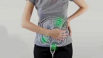
Diverticular disease is a common condition, especially in older adults. It involves the formation of small pouches or sacs (diverticula) that bulge through weak spots in the colon wall, primarily in the sigmoid colon. When these diverticula become inflamed or infected, acute diverticulitis develops , a potentially serious condition that requires accurate and prompt diagnosis.
Why is Ultrasound a Valuable Tool?
Accessibility and Speed: Ideal for initial assessment in emergency departments for abdominal pain.
Without Ionizing Radiation: Advantage over Computed Tomography (CT).
Dynamics: Allows evaluation of compressibility and pressure pain right at the point of interest (focused pain).
High Sensitivity: Modern studies show very high sensitivity and specificity for diverticulitis , especially in expert hands.
Key Ultrasound Findings:
A) In Simple Diverticular Disease (without acute inflammation):
Diverticula: These appear as small, rounded or oval structures protruding from the lumen of the colon. They may contain air ( hyperechoic with posterior "shadow cones") or fecal material ( echoic content ).
Intestinal Wall: Generally of normal thickness or slightly thickened symmetrically.
B) In Acute Diverticulitis :
Segmental and Asymmetric Wall Thickening: This is the cardinal finding. The colon wall is markedly thickened (often >4-5 mm) focally, usually in a long segment (>5 cm).
Pericolonic Fat Thickening (Panniculitis): The fatty tissue surrounding the inflamed colon becomes hyperechoic (whiter and "shinier") and edematous, losing its usual structure. It is a very sensitive sign.
Inflamed Diverticulum: This may appear as a bulging structure with a thickened, echogenic wall . Sometimes a small impacted fecal stone (fecalith) is identified in its neck, which appears hyperechoic .
"Omega" or "Communal Tooth" sign: In cross-section, the inflamed segment with diverticula may have a characteristic appearance.
Detectable Complications:
Pericolonic abscess : A fluid collection ( anechoic or hypoechoic ) with echogenic content (pus/ debris ), located adjacent to the inflamed colon. Doppler ultrasound may show peripheral enhancement.
Fistula: A linear hypoechoic tract connecting the colon to neighboring organs (bladder, vagina) or the skin. Air bubbles ( mobile hyperechoic spots ) may be seen in the bladder in cases of colovesical fistula .
Perforation with Free Air ( Pneumoperitoneum ): Although CT is more sensitive, on ultrasound free air can be seen as hyperechoic bands with reverberation artifacts ("streaks") that move with changes in position, under the diaphragm or between the liver lobes.
Advantages and Limitations:
Advantages: Dynamic, accessible, radiation-free, excellent for tracking collections.
Limitations: Operator-dependent, excessive intestinal gas can obstruct the view, and it is less sensitive for evaluating distant extensions or in patients with morbid obesity.
Conclusion:
Abdominal ultrasound is a first-line, effective, and safe tool for diagnosing acute diverticulitis and its complications. Its correct interpretation allows for timely medical treatment or guides percutaneous interventions in cases of abscesses, avoiding the need for studies involving ionizing radiation in many cases.
Ultrasound diagnosis diverticulitis
Dr. Jose Segnini, Radiologist / Diagnostic Medical Sonographer
MD Radiologist (Venezuela – Chile)
Board Certified Diagnostic Medical Sonographer (ARDMS, USA)
Mobile Ultrasound & Medical Supplies – Orlando, Florida


Comments